Electron microscope in action |
|||||||||
 In "Electron microscope in action" everybody can zoom in on the tiny little structures that can become visible with a FESEM (a 'Field-Emission Scanning Electron Microscope'). With this instrument details as small as only a few nanometers (1 nanometer = one millionth of a millimeter = 10-9m!) can be still be distinguished.
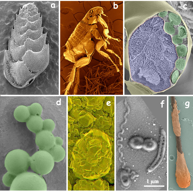
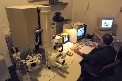
The FESEM shown here left (open zoom) is employed by scientists of the Radboud University to study a great variaty of objects, like cellular organelles, synthetical polymers and coatings on microchips. A number of such applications are demonstrated in this series of illustrated webpages. On each page a program can be operated which simulates the functioning of the FESEM on the object of choice (see options on the menu bar left). One can modify a number of variables like the magnification, the focus and the contrast in the same way as in the real microscope. (Link for help on how to operate the virtual FESEM). Like the name FESEM already indicates, this microscope does not work with light (photons) but with electrons. Electrons are generated in a 'source' (Emission) and accelarated under influence of a strong electrical voltage gradient (Field). With electromagnetic coils an electron beam is formed that scans the object (Scan) and secondary electrons are produced by interaction with the atoms at the surface of the sample. These electrons contain valuable information that is employed to reconstruct a very detailed image of the topography of the surface of the specimen (More explantions are given in the pages on the functioning of the FESEM and other electron microscopes). A few applications of the FESEM in research and teachingThe FESEM is een multipurpose instrument that is applied in diverse filed of research and education. See here below a few examples:
|
|||||||||