FESEM help |
||||
Download pdf help document (251 KB)
Help menu for the FESEM operation panelSee below for help on the operation panel of the virtual FESEM. The same help menu is also available as pdf document (251 KB) and as Word document (53 KB) for download.The Info - FESEM pages provide explanation on the functioning of the real FESEM.
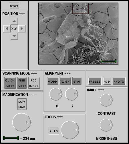
Manual operation panel of virtual FESEMFRAME. This frame displays the object that is positioned under the microscope and the changes in settings. The red frames, which are present in most images, indicate the region on which one can zoom in. The green scale bar is used to calibrate the magnification (see MAGNIFICATION).
POSITION X Y. The object under the microscope can be shifted horizontally and vertically on the screen by clicking on the arrows around X-Y. The duration of the pressure on the arrows determines the distance of the shift in the particular direction. The shift is only effected after release of the button. It is possible to zoom in to a higher magnification in the regions marked with a red frame, provided they are centered.
Reset. Clicking on reset puts all settings back to the initial situation.
SCANNING MODE
QUICK VIEW is virtually not active. In the real FESEM the function QUICK VIEW activates two scan modes: a rapid scan mode that is useful for observation while repositioning the object, and a standard scan mode that is employed to evaluate the effect of changes in settings. The scan speed correlates with the refreshing rate of the images and the noise level.
FINE VIEW Both the virtual and real FESEM produce a high quality image with FINE VIEW, however with a low scan speed. FINE VIEW is a useful function to observe details while scanning, but it is not appropriate to evaluate quick adjustments because of the low refreshing rate. THEREFORE, DO NOT TURN THE KNOBS TOO QUICKLY!
RDC means "reduced image". Only a small window in the center of the field is scanned. Because the area of this central region smaller than the full format, the refreshing rate is accordingly quicker. In the real FESEM, RDC scanning uses the same speed as the original full format mode. Switching from FINE VIEW to RDC, for example, results in a more rapid display of adjustments in a small area with the same sharpness as the original full image.
MAGNIFICATION. This knob regulates the magnification factor. The green bar under the knob shows how big (or how small!) the structures under the microscope are in reality. 1 �m corresponds to one thousand of a millimeter (0.001 mm), or 10 -6 m. By positioning the red frames in the center, it is possible to zoom in in these regions using the Magnification knob.
LOW MAG gives an overview image of the object.
ALIGNMENT: WOBB means wobbler, ALIGN alignment and STIG stigmator. By turning the X and Y knobs under ALIGNMENT a simulation can be seen of the aberrations in the image (i.e. astigmatism) caused by a poor alignment of the electron beam.
FOCUS is the knob to adjust the focus.
AUTO is virtually not active. In practice this knob activates a function that automatically corrects small focus deviations.
IMAGE CONTRAST BRIGHTNESS. These two knobs are for the adjustment of contrast and brightness in teh image. By clicking on ACB = Adjust Contrast en Brightness the two functions are automatically optimized.
FREEZE is virtually not active. In the real FESEM a number of scans can be averaged. The result is a sharp image with a strong signal and little noise.
PHOTO is virtually not active. In the real FESEM PHOTO switches a slow scan modus to built a high-quality digital image pixel by pixel. |
||||