Topics on this page:
- A. Upper and lower side of a leaf (More...)
- B. Typical anatomy of a leaf (More...)
- C. Cell types in leaves ( More...)
- C1. Normal and special epidermis cells (More...)
- C2. Mesophyll, chlorenchyma, palisade and spongy parenchyma (More...)
- C3. Vascular bundles: xylem and phloem (More...)
A. Upper and lower side of a leaf
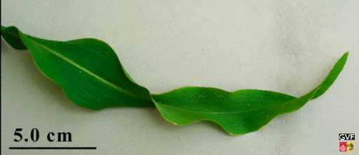
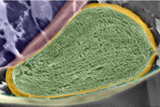
Question: Which side of this maize leaf is the true upperside?
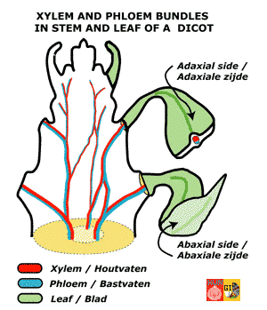
Which side of a leaf is the top side and which is the under side,is not always obvious. In general, the side oriented towards the sun bears an extra waxy -and shiny- protective layer: a -thicker- cuticula. Opposite, the lower side facing the shadow, thus the side where the risk of dessication is smaller, bears often relatively more tiny openings (stomata) for gas exchange. This side is often duller (see example of an ivy leaf). If you can dispose of a microscope, you could deduce where the anatomical upper and lower side lay from the orientation of the xylem and phloem vessels in the vascular bundles. The xylem vessels, recognizable as large and hollow tubular structures, are located toward the inner side in the stem, and they protrude to the anatomical upper side (= adaxial side) into the leaf. The phloem is directed to the outer margin in the vascular bundle of the stem, and they extend to the anatomical underside (= the abaxial side) of the leaf. The "adaxial" and "abaxial" side can thus be firmly determined independently of the position of the leaf relative to the sun, and therefore these terms are prevalent in botany.
|
|
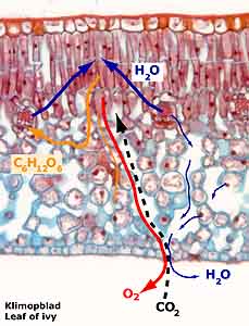
B. Typical anatomy of a leaf
All full grown leaves share a basic anatomy, due to their specialized function in photosynthesis. Below we describe the basic anatomy of the leaf using a cross section through the leaf of the ivy,
Hedera helix, as an example. The property common to most leaves that the adaxial side differs from
the abaxial side both in outer appearance and inner architecture, the so-called bifacial structure, can be clearly seen in ivy.
C. Cell types in leaves
C1. Normal and special epidermis cells
The epidermis is the outermost layer of the leaf, whicht serves to protect the inner tissues. It mostly consists of a single-cell layer, but sometimes multiple-layering can be encountered (see example here below in Ligustrum). In a transverse section of an entire leaf both upper end lower epidermis can be seen (see ivy here above). Often, the upper epidermis differs from the lower epidermis. Epidermal cells are mostly thick walled, they do not contain chloroplasts and they form an unbroken cover. The epidermis protects against a) dehydration, b) intense sunlight, especially ultraviolet (UV) irradiation, c) mechanical stress and d) predation by herbivores (insects, cattle). Among specific adaptations against animals, one can mention the occurance of
multi-layered epidermis with thick cell walls, hook-shaped and stingy
hairs and the presence of sticky, irritating, toxic or indigestible substances, like
tannins. Since dehydration is the major threat for plants, the epidermal cells are covered with a cuticle, a layer of cutin, a tough lipid-like and water impermeable substance. In addition a waxy layer is often present (See image of
wax deposition).
| Normal epidermal cells and hair cells |
|
|
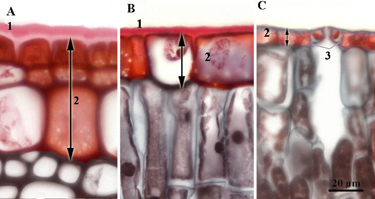
Upper epidermis of leaves in cross sections: A. Ligustrum, B. Ivy, C. Water lily:
1 Cuticula, 2 Upper epidermis, 3. Stomata (single cell layer in B and C, multiple in A).
Magnification in A, B, C is similar
|
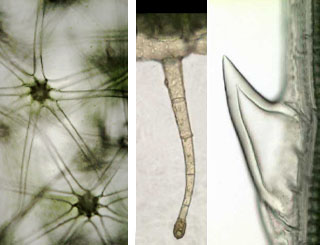
Leaf hairs in Solanum quadriloculatum, Petunia and Papyrus:
Examples of leaf hairs providing protection against resp. dessication, damage by bugs (secretion of sticky substance) and hog (sharp hooks)
|
A special adaptation to land-life is the occurrence of
stomata that can be actively opened or closed and thus allow a controlled gas exchange with the environment. Submerged plants do not possess stomata; gas exchange occurs via the epidermis via a thin air admissible cuticle.
Opening and closing of the stomata by the paired
guard cells controls the gas exchange rate. In some cases, the guard cells are
supported by the
subsidiary cells. Guard cells act as ports between the environment and the interior of the leaf. When the guard cells accumulate water they become turgescent and open. Opening and closing in turn is controlled by light conditions, air humidity, temperature and the CO
2 concentration. Guard cells, in contrast to other epidermis cells, do possess chloroplasts.
| Stomata: opening, guard cells and subsidiary cells |
 |
|
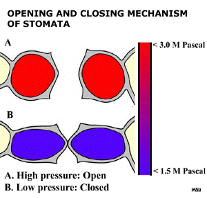
 Stomata open when the internal pressure of the guard cells rises as a result of water absorption. The pressure increases from 1.5 to 3.0 mega Pascal. Since the walls of the guard cells are relatively flexible at the side of the stoma, the guard cells expand vertically and the stoma subsequentely opens.
Click on this gif animation to download a larger film (about 260 Kb) on this mechanism.
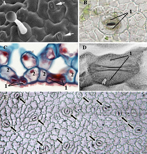
Right. Stomata: in SEM (A), upperview of fresh leaf material in bright field microscopy (B), in a stained cross-section viewed with light microscopy(C), in a SEM close-up (D) and impression of stomata and epidermal cells on a nail polish film (E)
1 Guard cells, 2 Subsidiary cells
|
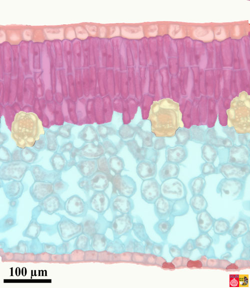
C2. Mesophyll, chlorenchyma, palisade and spongy parenchyma
The tissue specialized in photosynthesis is the mesophyll or chlorenchyma. The mesophyll commonly is differentiated in palisade and spongy parenchyma.
| Mesophyll |

|

|
Palissade and sponge parenchyma in a fresh piece of leaf of Petunia:
1 upper epidermis, 2 palisadeparenchyma, 3 spongy parenchyma, 4 air cavity
zoom
|
Scanning electron microscopy image of a cross-section through a leaf of Petunia (colored):
1 upper epidermis, 2 palisadeparenchyma, 3 spongy parenchyma, 4 air cavity, 5 lower epidermis
zoom
|
|
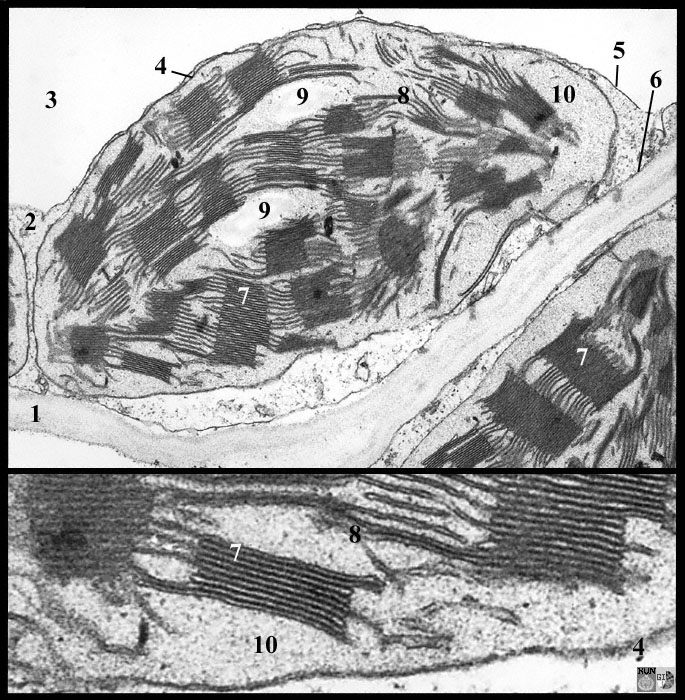
Chloroplast viewed in transmission electron microscopy (left in a tobacco leaf; zoom) and in a scanning electron microscope (here below, in petunia; colored image):
1 cell wall, 2 cytoplasm, 3 vacuole, 4 chloroplast envelope (2 membranes), 5 tonoplast, 6 plasma membrane, 7 grana, 8 stroma thylakoids, 9 starch grains, 10 stroma
|
|
The palisade (pole-shaped) parenchyma is located directly under the upper epidermis. The cylindrical and elongated cells (or palisades) are at right angles to the epidermis and they contain chloroplasts (see part on
photosynthesis). The palisade parenchyma is the main photosynthetic tissue of the leaf.
The spongy parenchyma has an open and net-like structure with large inter-cellular spaces that facilitate gas diffusion. The spongy parenchyma also contains
chloroplasts and contributes to photosynthesis. It therefore is part of the chlorenchyma. The major function of the spongy parenchyma is the transport of oxygen, carbon-dioxyde and water vapour. It also is involved in the transport of water and the products of photosynthesis, the sugars. The spongy parenchyma is in close connection with the vascular bundles and the palisade parenchyma. When no clear differentiation exists between palisade and spongy parenchyma, the tissue is called mesophyll.
C3. Vascular bundles: xylem and phloem
The vascular bundles, or veins, of the leaf are irregularly distributed throughout the mesophyll.
They transport water taken up in the roots through the xylem to the leaf and for the distribution of sugars, as products of photosynthesis, from the leaf through the phloem to all the parts of the plant.
To a page on this site on the electron microscopy of leaf veins.
| Vascular bundles |
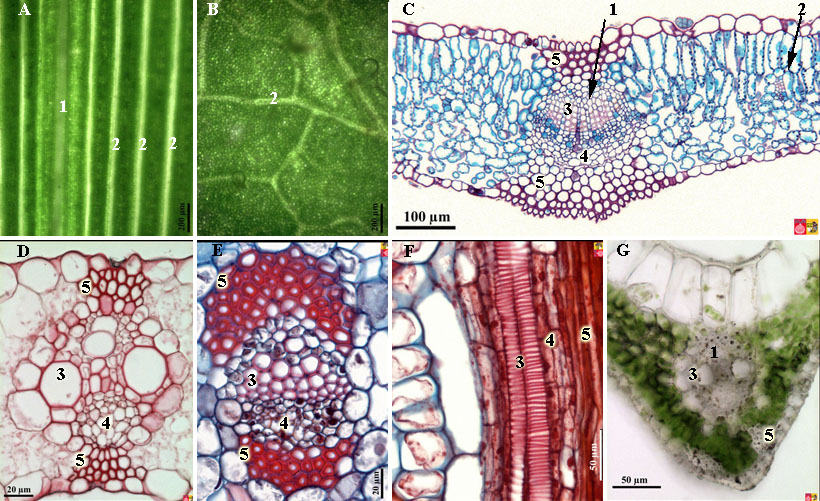 |
A. Upperview of a leaf of papyrus (monocot) and B. ivy (dicot). C. Cross-section through a leaf of lilac. D. Cross-section through a main vascular bundle of maize and E. Yucca. F. Longitudinal section through a vascular bundle of the waterlily and G. Fresh section through a main vein of Papyrus.
1 Main vein, 2 Secondary veins, 3. Xylem, 4 Phloem, 5 Sclerenchyma |


