Chloroplasts in sponge parenchyma of leaves: the lightcatchers
| To the fesem simulator... |
|
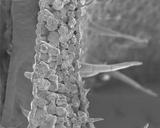
Click on the gray image here left to load the virtual FESEM or
open a new window.
The FESEM simulator works with Java. If the required Java (TM) plug-in 1.3 is not installed yet on your computer, you will be automatically redirected to Sun Microsystems, Inc. Follow the (simple) step-by-step instructions to download the free plug-in. After completion of the installation procedure the virtual FESEM will be launched automatically.
|  |
Sponge parenchyma and chloroplast
Plants utilize their leaves to produce sugars in a process called photosynthesis. Photosynthesis starts with the capture of light energy from the sun by pigments that are located in the internal membranes of chloroplasts. Chloroplasts are abundant in the sponge parenchyma of leaves.
With field-emission scanning electron microscopy (FESEM) and transmission electron microscopy (TEM) very clear details of the structure of chroroplasts can be observed.
| FESEM views of sponge parenchyma with chloroplasts |
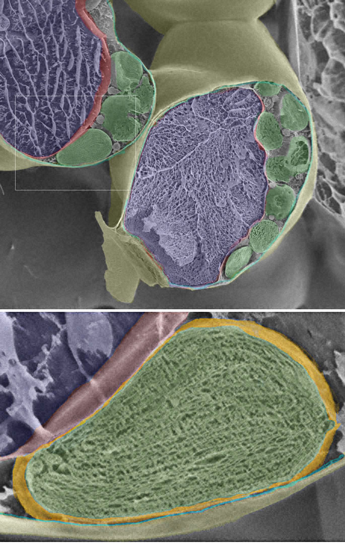
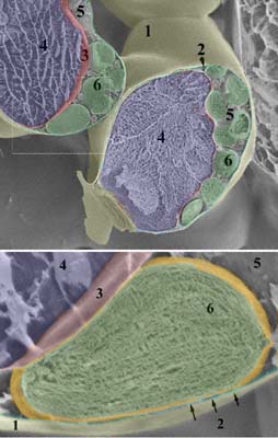 | Overview (original image is in gray shades; structures indicated here by color overlays): This view shows the cell wall that surrounds the cell content (1; yellow). The plasma membrane (2 and arrows; blue) is appressed along the cell wall. The tonoplast (3; pink) ensheathes the vacuole (4; purple). The cytoplasm (5) occupies the space between the plasma membrane and the tonoplast. In the cytoplasm distinction is made between the cytosol (fluid) and organelles of various types, among which chloroplasts (6; green), mitochondria, endoplasmic reticulum, Golgi (not shown). The cell nucleus is not visible in this fracture.
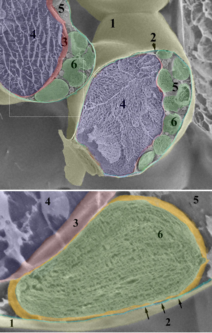
Zoom image of a single chloroplast: Each chloroplast (6) has an own envelop (painted orange here) that consists of two membranes. The inner compartment of the chloroplast (green) houses a complex system of thylakoid membranes that are differentiated into lamellae and stacks of disks called grana. The space around the thylakoids is named stroma. The thylakoids are the sites where the photochemical reactions of photosynthesis occur. The stroma is the site of the biochemical carbon reduction reaction.
|
Material: a piece fresh Coleus leaf material was frozen with slush nitrogen at - 95 ° C. In order to observe the inner part of single cells, this small piece of tissue was fractured using with a special knife in the cryo-unit of the FESEM.
High-resolution SEM image of the sponge paremchyma cells and the chloroplast © with labels or without labels (172 KB). Printable version of this page in Word or pdf format.
|
|
| TEM view of a single chloroplast and details | 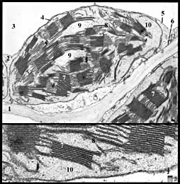 | Chloroplast of tobacco:
1 = cell wall
2 = cytoplasm
3 = vacuole
4 = chloroplast envelope (2 membranes)
5 = tonoplast
6 = plasma membrane
7 = grana
8 = stroma thylakoids
9 = starch grains
10 = stroma
More information on photosynthesis is available in the virtual lesson on leaves. |
Software development: Jeroen van Beurden. Web structure: Remco Aalbers. Text and images: Liesbeth Pierson and Huub Geurts