 |
Electron microscopy (EM) |
Further information and images on preparation in TEM
|
Electron microscopy (EM) allows one to visualize objects that are as small as 1 nm (one nanometer is equal to one thousand of a micrometer, is one million of a millimeter or one billion of a meter). To produce an image the sample is bombarded by a beam of electrons.
Globally, distinction is made between two EM techniques:
- Transmission Electron Microscopy (TEM). TEM allows one the study of the inner structure and contours of objects (tissues, cells, virusses)
- Scanning Electron Microscopy (SEM). SEM is applied to visualize the surface of tissues, macromolecular aggregates and materials.
More on how an EM works...
| 
To the virtual FESEM simulator and various examples of applications of a SEM.
|
| Examples of TEM and SEM images |
|
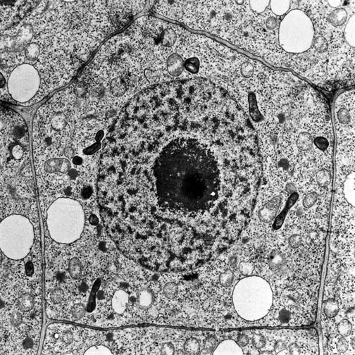
TEM allows one to capture high-resolution (± 1 nm) views of the inner structure of objects, like cells, or of the total structure of ultrathin material (< 1 µm, like virusses and aggregates of macromolecules). TEM usually requires cuts of the material in extremely thin sections (this is the case for most cells) and 'staining' with heavy metals before visualization.
|
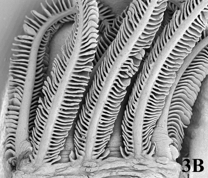
In SEM an image of the surface of the object is created. That image is formed by secondary electrons which are liberated by the electron beam that bombards the object.
|
 |
 |
Plant cell (root tip cell) imaged with a Transmission Electron Microscope (TEM)
Imaging: A.M. Wolters-Arts, Radboud University Nijmegen. | Gills of a fish, the mudskipper (Periophthalmus argentilineatus) taken with a Scanning Electron Microscope (SEM)
Imaging: G. Kruitwagen and H.P.M. Geurts, Radboud University Nijmegen. |
last modified: 26 Nov 2010 |
 |
|
|


